Parkinson´s Disease


Parkinson's ( or Parkinson´s disease or PD ) is a degenerative disorder of the central nervous system that often impairs the sufferer's motor skills and speech, as well as other functions. It is a progressive disease whose effects get worse over time.

PD is the most common cause of chronic progressive parkinsonism, a term which refers to the syndrome of tremor, rigidity, bradykinesia ( slow movement ) and postural instability. While many forms of parkinsonism are "idiopathic" meaning that they are not caused by any other agent, secondary cases may result from toxicity most notably of drugs, head trauma, or other medical disorders


Symptoms of Parkinson's disease

People who have Parkinson's disease experience the following symptoms:

1. Tremor. The characteristic shaking associated with Parkinson's disease often begins in a hand. However, many people with Parkinson's disease do not experience substantial tremor.

2. Slowed motion (bradykinesia). Over time, Parkinson's disease may reduce your ability to initiate voluntary movement. This may make even the simplest tasks difficult and time-consuming. When you walk, your steps may become short and shuffling.

3. Rigid muscles. Muscle stiffness often occurs in your limbs and neck. Sometimes the stiffness can be so severe that it limits the range of your movements and causes pain.

4. Impaired posture and balance. Your posture may become stooped as a result of Parkinson's disease. Imbalance also is common, although this is usually mild until the later stages of the disease.

5. Loss of automatic movements. Blinking, smiling and swinging your arms when you walk are all unconscious acts that are a normal part of being human. In Parkinson's disease, these acts tend to be diminished and even lost.

6. Speech changes. Many people with Parkinson's disease have problems with speech. You may speak more softly, rapidly or in a monotone, sometimes slurring or repeating words, or hesitating before speaking.

7. Dementia. In the later stages of Parkinson's disease, some people develop problems with memory and mental clarity. Alzheimer's drugs appear to alleviate some of these symptoms to a mild degree.


Diagnosis

A doctor may diagnose a person with Parkinson's disease based on the patient's symptoms and medical history. The disease can be difficult to diagnose accurately. The Unified Parkinson's Disease Rating Scale is the primary clinical tool used to assist in diagnosis and determine severity of PD. It consists of the following sections:

Mentation, behavior, and mood;
Activities of daily living;
Motor;
Complications of therapy;
Hoehn and Yahr Stage;
Schwab and England Activities of Daily Living Scale
These are evaluated by interview and clinical observation. Some sections require multiple grades assigned to each extremity.


What causes Parkinson's disease?

Many symptoms of Parkinson's disease result from the lack of a chemical messenger, called dopamine, in the brain. This occurs when the specific brain cells that produce dopamine die or become impaired. But researchers still aren't certain about what sets this chain of events in motion. Some theorize that genetic mutations or environmental toxins may play a role in Parkinson's disease.


Treatment

Parkinson's disease is a chronic disorder that requires broad-based management including patient and family education, support group services, general wellness maintenance, physiotherapy, exercise, and nutrition. At present, there is no cure for PD, but medications or surgery can provide relief from the symptoms.

Some of the medicines used to treat Parkinson's disease include carbidopa-levodopa (one brand name: Sinemet), bromocriptine (brand name: Parlodel), selegiline (one brand name: Eldepryl), pramipexole (brand name: Mirapex), ropinirole (brand name: Requip), and tolcapone (brand name: Tasmar). Your doctor can recommend the best treatment for you.

Alternative Medicine

Diet

The choices we make about food – what we consume, its quality and quantity – arecrucial to our health and well-being. There is some agreement that it is generally wise to consume a varied diet high in fruits and vegetables and to avoid excessive saturated fats, especially trans-fats. For example, Mediterranean diet, a diet high in monounsaturated fats, such as olive oil, may be beneficial in reducing blood pressure and cardiovascular disease. The diet also emphasizes fish, especially those high in Omega-3 fatty acids, such as salmon, and foods containing antioxidants.

Antioxidants

Antioxidants are substances that can detoxify free radicals, which are reactive particles involved in certain types of cell death. Coenzime Q10 is one antioxidant that lately has appeared to have some effect on disease progression at doses of 1200mg/day. This is a much larger dose than most people were taking prior to the release of the study data, a deliberate choice designed to avoid the risk of missing a significant effect by giving too low a dose. Another medications with far less research include IV Gluthathione and very high dose Vitamin C.

Ayurvedic Medicine

The initial step is to determine the metabolic type of an individual. Then the practitioner looks at environmental factors, such as season and time of day. Diseases are diagnosed by assessing various pulse points and their relationship to internal organs.

Treatment of disease consists of detoxification through various cleansing therapies, then restoring balance through palliation with such modalities as yoga and meditation. Finally, a process of tonification, called rasayana, is initiated. Interestingly, one of the medications used in Ayurveda was derived from a legume, Mucuna pruriens, which has been found to contain levodopa. The condition for which it was used, as described many centuries ago, most likely was Parkinson disease.

Yoga

A complete practice of yoga integrates mind, body and spirit in a process involving one’s complete lifestyle. The most popular form of yoga is asana or Hatha yoga, which involves execution of a series of postures with attention to breathing (pranayama), meditation and proper execution of the poses. Like Tai Chi, the practice of yoga has been shown to improve various aspects of health such as blood pressure, digestion and asthma. Most yoga centers offer a range of classes and list them as to level of experience required.

Since exercise is so important in the treatment of PD, patients are seeking to improve their strength, balance and flexibility. Yoga and Tai Chi are both excellent for this. In addition to more traditional fitness programs such as walking and weight lifting, classes in yoga and Tai Chi have become standard offerings in senior centers, gyms and park districts.

Traditional Chinese Medicine (TCM)

Like Ayurveda, Chinese or Oriental Medicine has been in practice for thousands of years and also is concerned with maintaining health instead of just reacting to disease. Much emphasis is placed on maintaining a balance between opposites within the body as well as with the natural world.

Tai Chi

Tai Chi, both a form of martial arts and a system of meditation, is part of an ancient Chinese system of healing called Qigong. In Tai Chi classes, participants follow a teacher in performing a choreographed series of movements. There are various styles and levels of difficulty, including some classes performed while practitioners are seated. Tai Chi has been shown in several studies to improve balance in older patients as well as persons with PD.

Acupuncture

One of the methods used to help restore the balance of yin and yang is acupuncture, a technique developed over 2,500 years ago in China. The treatment involves inserting hair-like needles into certain points on the body. This is done to restore the flow of Qi to the organ system associated with that acupoint. Some patients with PD describe temporary relief from symptoms such as tremor and rigidity. Variants of acupuncture include cranial acupuncture and seem to be effective in helping PD patients.

Body Work/Massage Therapy

Body work comprises a group of “touch therapies” such as reflexology, and therapeutic massage. Massage therapy in particular has become very popular because of its beneficial effects on the muscle stiffness and aching that may accompany PD. Massage may also help with associated conditions such as arthritis, and sleep and digestive disorders. In addition, a well-executed massage can be an extremely relaxing and enjoyable experience.

Of the many different styles of massage therapy, two in particular may be useful in PD. Shiatsu, or acupressure, uses touch rather than needles to treat the same pressure points as acupuncture.


Please look at the following video

Seasonal Affective Disorder


Seasonal affective disorder (also called SAD) is a type of depression that follows the seasons and is usually called winter depression. It usually begins in late fall or early winter and goes away by summer. SAD may be related to changes in the amount of daylight during different times of the year.


With respect to prevalence, about half a million people in the United States may have winter depression. Another 10% to 20% may experience mild SAD. SAD is more common in women than in men. For adults, the risk of SAD decreases as they get older. SAD is more common in northern geographic regions, where the exposure to bright light is obviously reduced during winter season.


Why does seasonal affective disorder occur?


Seasonal mood variations are believed to be related to light. An argument for this view is the effectiveness of bright light therapy. SAD is measurably present at latitudes in the Arctic region, such as Finland (64º 00´N) where the rate of SAD is 9.5%. Cloud cover may also contribute to the negative effects of SAD.


SAD can be a serious disorder and may require hospitalization. There is also potential risk of suicide in some patients experiencing SAD. One study reports 6-35% of sufferers required hospitalization during one period of illness. The symptoms of SAD mimic those of clinical depression. At times, patients may not feel depressed, but rather have depression equivalent symptoms such as a lack energy to perform everyday activities. Norman Rosenthal, a pioneer in SAD research, has estimated that the prevalence of SAD in the adult United States population is between about 1.5 percent in Florida and about 9 percent in the northern US.


Various causes have been suggested. One possibility is that SAD is related to a lack of serotonin, although this theory has not been widely accepted.


How does my doctor know I have SAD?


1. Appetite changes, especially a craving for sweet or starchy foods
2. Weight gain
3. Heavyness in the arms or legs
4. Decreased energy and fatigue
5. Oversleep need
6. Difficulty concentrating
7. Irritability or excessive passiveness
8. Increased sensitivity to social rejection


According to the American Psychiatric Association, for a diagnosis to qualify as SAD, it must meet four criteria:


I. Depressive episodes at a particular time of the year


II. Remissions or mania/hypomania also at a characteristic time of year


III. These patterns must have lasted two years with no nonseasonal major depressive episodes during that same period


IV. The seasonal depressive episodes outnumber other depressive episodes throughout the patient's lifetime.


Treatment for SAD


Winter depression is probably caused by your body's reaction to a lack of sunlight. Light therapy is one option for treating winter depression. You will need to sit in front of the light box or wear the light visor for a certain length of time each day. Generally, light therapy takes about 30 minutes each day throughout the fall and winter, when you're most likely to be depressed. The sufferer sits a prescribed distance, commonly 30-60 cm, in front of the box with her/his eyes open but not staring at the light source. Many patients use the light box in the morning, however it has not been proven any more effective than any other time of day. Discovering the best schedule is essential because up to 69% of patients find it inconvenient. Dawn simulation has also proven to be more effective in some studies, there is an 83% better response when compared to other bright light therapy


When used properly, light therapy seems to have very few side effects. Side effects may include eyestrain, headache, fatigue, irritability and inability to sleep (if light therapy is used too late in the day).


Tanning beds shouldn't be used to treat SAD. The light sources in tanning beds are high in ultraviolet (UV) rays, which harm both your eyes and your skin.


In addition to the light box your doctor may also want you to try a medicine or behavior therapy to treat your SAD. If light therapy or medicine alone doesn't work, your doctor may want you to use them together.


Please view the following video related to SAD

Plantar Fasciitis


Are your first steps out of bed in the morning causing you severe pain in your heel? Or does your heel hurt after jogging or playing tennis?


Most commonly, heel pain is caused by inflammation of the plantar fascia which is a tendon, that starts at your heel and goes along the bottom of your foot. It attaches to each one of the bones that form the ball of your foot. The plantar fascia works like a rubber band between the heel and the ball of your foot to form the arch of your foot. If the band is short, you'll have a high arch, and if it's long, you'll have a low arch, what some people call flatfeet. A pad of fat in your heel covers the plantar fascia to help absorb the shock of walking. Damage to the plantar fascia can be a cause of heel pain. The condition is called plantar fasciitis


Plantar fasciitis causes stabbing or burning pain that's usually worse in the morning because the fascia tightens (contracts) overnight. Once your foot limbers up, the pain of plantar fasciitis normally decreases, but it may return after long periods of standing or after getting up from a seated position.


Risk Factors and Causes


When we age, the plantar fascia becomes less like a rubber band and more like a rope ( stiffens up ) and is unable to stretch very well. The fat pad on the heel becomes thinner and can't absorb as much of the shock caused by walking. What happens next is that plantar fascia may swell, tear or bruise. You may notice a bruise or swelling on your heel.


1. Physical activity overload. Plantar fasciitis is common in long-distance runners. Jogging, walking or stair climbing also can place too much stress on your heel bone and the soft tissue attached to it, especially as part of an aggressive new training regimen.
2. Arthritis. Some types of arthritis can cause inflammation in the tendons in the bottom of your foot, which may lead to plantar fasciitis.
3. Diabetes. Plantar fasciitis occurs more often in people with diabetes.
4. Faulty foot mechanics. Being flat-footed, having a high arch or even having an abnormal pattern of walking can adversely affect the way weight is distributed when you're on your feet, putting added stress on the plantar fascia.
5. Use of improper shoes. Shoes that are thin-soled ( as flip-flops do ) , loose, or lack arch support or the ability to absorb shock don't protect your feet. If you regularly wear shoes with high heels, your Achilles tendon — which is attached to your heel — can contract and shorten, causing strain 6. Overweight and obesity
7. Spending most of the day on your feet


Signs and symptoms

Plantar fasciitis usually develops gradually, but it can come on suddenly and be severe. And although it can affect both feet, it more often occurs in only one foot at a time. Signs and symptoms include:


1. A sharp or knife-like pain in the inside part of the bottom of your heel or bottom of foot.


2. Heel pain that tends to be worse with the first few steps after awakening, when climbing stairs or when standing on tiptoes.
3. Heel pain after long periods of standing or after getting up from a seated position
4. Heel pain after, but not usually during, exercise
5. Swelling in your heel


Treatment


It typically takes six to eighteen months to find a favorable resolution to plantar fasciitis, but it has a generally good long-term prognosis.


1. Stretching Exercises


Stretching exercises ( need to be done at home constantly ). The mainstays of treatment are stretching the Achilles tendon and plantar fascia, resting, keeping off the foot as much as possible, discontinuing aggravating activity. Common sense recommendations include wearing supportive and stable shoes. Patients should avoid open-back shoes, sandals, "flip-flops", and any shoes without a raised heel. Two of the preferred exercises that are usually recommended are:


A. Towel stretch: Sit on a hard surface with your injured leg stretched out in front of you. Loop a towel around the ball of your foot and pull the towel toward your body keeping your knee straight. Hold this position for 15 to 30 seconds then relax. Repeat 3 times.
When the towel stretch becomes to easy, you may begin doing the standing calf stretch.


B. Frozen can roll: Roll your bare injured foot back and forth from your heel to your mid-arch over a frozen juice can. Repeat for 3 to 5 minutes. This exercise is particularly helpful if done first thing in the morning.


C. Towel pickup: With your heel on the ground, pick up a towel with your toes. Release. Repeat 10 to 20 times. When this gets easy, add more resistance by placing a book or small weight on the towel.


2. Physical Therapy Modalities:


A physical therapist can instruct you in exercises to stretch the plantar fascia and Achilles tendon and to strengthen lower leg muscles, which stabilize your ankle and heel. A therapist may also teach you to apply athletic taping to support the bottom of your foot. Modalities include cold compression therapy, contrast bath therapy, iontophoresis and weight loss.


3. Orthotics.


Your doctor may prescribe off-the-shelf or custom-fitted arch supports (orthotics) to help distribute pressure to your feet more evenly.


4. Night splints.


Your doctor may recommend wearing a splint fitted to your calf and foot while you sleep. This holds the plantar fascia and Achilles tendon in a lengthened position overnight so that they can be stretched more effectively.


5. Oral Medications:


To relieve pain and inflammation, nonsteroidal anti-inflammatory drugs (NSAIDs) such as aspirin and ibuprofen are often used but are of limited benefit. Patients should be encouraged to lessen activities which place more pressure on the balls of their feet because it increases tension in the plantar fascia.


6. Corticosteroid Injection at the site of pain.


This modality often gives temporary or permanent relief, but may be painful, especially if not combined with a local anesthetic and injected slowly with a small-diameter needle. Repeated steroid injections may result in rupture of the plantar fascia.


7. Therapeutic ultrasound


It has been shown in a controlled study to be ineffective as a treatment for plantar fasciitis. More recently, however, extracorporeal shockwave therapy (ESWT) has been used with some success in patients with symptoms lasting more than 6 months.


Most patients ( over 95% ) should improve within one year of beginning non-surgical treatment, without any long-term problems. A few patients, however, will require surgery.


8. Surgery


Surgical procedures, such as plantar fascia release, are a last resort, and often lead to further complications such as a lowering of the arch and pain in the supero-lateral side of the foot due to compression of the cuboid bone. The most common procedure is a partial plantar fasciotomy that may be either open or closed. An open procedure requires a 3-6 cm plantar medial incision to release the fascia. Nerve decompression and/or resection of calcaneal spur may also be performed at this time. A closed procedure utilizes endoscopy to release the fascia. An ultrasound guided needle fasciotomy can be used as a minimally invasive surgical intervention for Plantar Fasciitis. A needle is inserted into the Plantar Fascia and moved back and forwards to disrupt the fibrous tissue.


Alternative Medicine


a. Magnetic Insoles


Use of magnetic insoles has been considered by some clinicians as a treatment for PF, but available data regarding efficacy is limited, and results are mixed at best.


b. Acupuncture


Acupuncture has been proposed as an effective treatment for PF


c. Nutritional Considerations


-Vitamin C


Widely known for its antioxidant properties, vitamin C is also an essential component for healthy connective tissue repair. A deficiency in vitamin C can result in abnormal collagen fibers, as well as other changes in the intracellular matrix, that can contribute to decreased tensile strength of fibrous tissues, such as those found in the plantar fascia. Recommended dosing of vitamin C for the purpose of tissue repair is 1-3 g daily until resolution.


-Zinc


Zinc, an essential trace mineral, is utilized in over 300 known enzymatic reactions. It is a key element in tissue regeneration and repair, working in concert with vitamin C to increase tensile strength of wounded tissue.


Recommended dosage for zinc is 15-30 mg daily. (47,48)


-Glucosamine


Glucosamine may be helpful in the management of PF because it serves as a potential alternative to NSAID use and is a key biochemical component in the repair and regeneration of connective tissue. Glucosamine is the foundational structure of many compounds associated with repair and regeneration of connective tissue. It is the essential substrate for hyaluronic acid and other glycosaminoglycans used in maintaining healthy joint function. In vitro studies suggest glucosamine stimulates the synthesis of glycosaminoglycans and collagen. Recommended dosage for glucosamine is 500 mg three times daily in the form of glucosamine sulfate.


-Bromelain


Bromelain is the singular name used for a family of proteolytic enzymes found in the pineapple plant. Bromelain is commonly used in treating inflammation and soft tissue injuries and, as such, may be beneficial in the management of PF. It has been shown to accelerate healing from bruises and hematomas. It was reported that use of bromelain reduced swelling, tenderness, and pain, both at rest and during movement.


There are several designations used to indicate the activity of bromelain. The most common measures of bromelain activity are milk clotting units (mcu) and gelatin dissolving units (gdu). One gdu approximately equals 1.5 mcu. Typically, enzymatic activity is given as a measure of mcu or gdu per gram of bromelain; e.g., 2,000 mcu/g or 1,333 gdu/g, respectively


-Fish Oil


These fish oils have been observed to suppress production of inflammatory mediators in patients with autoimmune conditions, such as rheumatoid arthritis. This may be due to reduced synthesis of key inflammatory mediators--leukotrienes, interleukin-2, and tumor necrosis factor. Such anti-inflammatory properties may in turn be beneficial in the management of PF. Recommended dosage for PF is 2-3 g daily.


Homeopathy


Homeopathy wise, consider the use of Bryonia 30C and Arnica 30C 4 pillules 3-4 times a day.


Deep Venous Thrombosis

What is deep vein thrombosis?

Deep vein thrombosis (also called DVT) is a blood clot in a vein deep inside your body. These clots usually occur in one of the major deep veins of the lower legs, thighs, or pelvis. A clot blocks blood circulation through these veins, which carry blood from the lower body back to the heart. The blockage can cause pain, swelling, or warmth in the affected leg.your leg veins. While DVT is a fairly common condition, it is also a dangerous one. Occasionally the veins of the arm are affected (known as Paget-Schrötter disease). If tlood clot breaks away and travels through your bloodstream, it could block a blood vessel in your lungs. This blockage (called a pulmonary embolism) can be fatal. Statistics reveal that at least 650,000 patients die each year from pulmonary embolism, making it the third most common cause of death in the United States.


In the United States, about 2 million people per year develop DVT. Most of them are aged 40 years or older. Up to 600,000 are hospitalized each year for the condition.

View the following animation about DVT and Pulmonary embolus


Signs and symptoms

There may be no symptoms referrable to the location of the DVT ( 30-50% of the patients will experience no symptoms ), but the classical symptoms of DVT include pain, swelling and redness of the leg, dilation of the surface veins, gradual onset of pain, warmth to the touch, worsening leg pain when bending the foot, leg cramps, especially at night and/or bluish or whitish discoloration of skin.

A careful history has to be taken considering risk factors (see below ). On physical exam, several techniques can be used for the detection of DVT, such as measuring the circumference of the affected and the contralateral limb at a fixed point (to objectivate edema), and palpating the leg veins, which is often tender. Physical examination is unreliable for excluding the diagnosis of deep vein thrombosis.

Causes of DVT

Three factors may lead to formation of a clot inside a blood vessel.

1. Damage to the inside of a blood vessel due to trauma or other conditions
2. Changes in normal blood flow, including unusual turbulence, or partial or complete blockage of blood flow
3. Hypercoagulability, a rare state in which the blood is more likely than usual to clot ( your physician should order blood tests to diagnose a primary hypercoagulability state ).

Risk Factors of DVT

Prolonged sitting, such as during a long plane or car ride
Prolonged bed rest or lack of movement
Recent surgery, particularly orthopedic, gynecologic, or heart surgery
Recent trauma or fractured bones of the hip, thigh, or lower leg
Obese patients
Recent childbirth
High altitude ( Over 4000 masl )
Use of estrogen replacement (hormone therapy, or HT) or birth control pills
Cancer
Genetic changes in certain blood clotting factors
Disseminated intravascular coagulation (DIC)
Advanced age

A second DVT is much more likely to happen after a first one.

Diagnosis of DVT

Physical exam will consist of:

A. Homan's test: Dorsiflexion of foot elicits pain in posterior calf. Rarely done due to its little diagnostic value and is theoretically dangerous because of the possibility of dislodgement of loose clot.
B. Pratt's sign: Squeezing of posterior calf elicits pain.

These two medical signs do not perform well and are subjective. Lately clinical prediction rules that combine best findings are used to diagnose DVT. Scarvelis and Wells overviewed a set of clinical prediction rules for DVT in 2006.

Blood tests

Tests such as complete blood count, coagulation studies ( PT, APTT ), fibrinogen, liver enzymes, renal function and electrolytes are regularly ordered. Also a test know as D-dimer is performed. This cross-linked fibrin degradation product is an indication that thrombosis ( formation of a clot ) is occurring, and that the blood clot is being dissolved by plasmin. A negative D-dimer is definitive to exclude the possibility of a blood clot.

Imaging studies

The gold standard is intravenous venography, which involves injecting a peripheral vein of the affected limb with a contrast agent and taking X-rays, to reveal whether the venous supply has been obstructed. Because of its invasiveness, this test is rarely performed. Lately doppler ultrasonography which is a compression ultrasound scanning of the leg veins, combined with duplex measurements (to determine blood flow), can reveal a blood clot and its extent (i.e. whether it is below or above the knee). It has got high sensitivity, specificity and reproducibility.

Treatment

A. At home

To increase comfort and lower the risk of the clot moving to the lung, the patient needs to keep the affected limb elevated, avoid prolonged sitting or bed rest and needs to use warm and moist heat to the area to relieve pain and inflammation.

Elastic compression stockings should be routinely applied beginning within 1 month of diagnosis of proximal DVT and continuing for a minimum of 1 year after diagnosis.

B. Medications

Anticoagulation is the usual treatment for DVT. In general, patients are initiated on a brief course (i.e., less than a week) of heparin ( fractionated or unfractionated ) treatment while they start on a 3- to 6-month course of warfarin.

C. Filter

If the patient can't take warfarin, or if a blood thinner doesn't work, your doctor may recommend that a filter is put into the vena cava (the main vein going back to your heart from your lower body). This filter can catch a clot as it moves through your bloodstream and prevent it from reaching your lungs. This treatment is used mostly for people who have had several blood clots travel to their lungs.


Hospital inpatient treatment is considered for patients with more than two of the following risk factors as these patients may have more risk of complications during treatment: bilateral DVT, renal insufficiency and cancer.


Diverticulosis and Diverticulitis


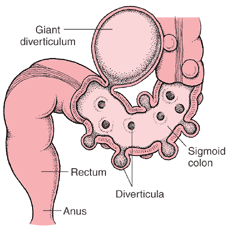
Diverticular disease affects the lining of the bowel. It is caused by small pouches (called diverticula) that can form anywhere in your digestive tract. They occur when the inner layer of the digestive tract bulges through weak spots in the outer layer.

Although these pouches can occur anyplace from the mouth to the anus, most occur in the large intestine (colon), especially the left (lower) part of the colon just above the rectum. These marble-sized pouches usually occur where blood vessels run through the intestinal wall. People who have these pouches are said to have diverticulosis. When this condition does not cause any symptoms, most people are unaware that they have it.

Sometimes when one or more of these pouches becomes inflamed or infected, a condition called diverticulitis is developed. Some people with diverticulosis become aware of the condition only when diverticulitis occurs. Diverticulosis is a very common condition in the United States. Diverticulosis is more common in developed or industrialized countries. In places such as the United States, England, and Australia, where the typical diet is low in fiber and high in highly processed carbohydrates, diverticulosis is common.

Diverticulosis first appeared in the United States in about 1900. This was the same time that processed foods were first introduced into the US diet.

Diverticulosis is much less common in countries of Asia and Africa, where the typical diet is high in fiber.

Most people recover from diverticulitis without problems if they receive appropriate treatment. Diverticulosis and diverticulitis can be prevented by changes in lifestyle and habits.

Causes

Diverticula are thought to be caused by increased pressure within the lumen ( inner aspect ) of the colon. Increased intra-colonic pressure secondary to constipation may lead to weaknesses in the colon walls giving way to diverticula. Fiber causes stools to retain more water and become easier to pass (either soluble or insoluble fiber will do this). A diet without sufficient fiber makes the stools small, requiring the bowel to squeeze harder to remove the smaller stool, thus increasing pressure.

Risk factors

1. Low fiber or high fat diets
2. Elderly
3. Constipation
4. Connective tissue disorders ( cause weakness in the colon wall such as Marfan syndrome).

Symptoms

Diverticulosis has almost no symptoms. The most common is bleeding (variable amounts), bloating, abdominal pain/cramping after meals or otherwise often in the left lower abdomen, and changes in bowel movements (diarrhea or constipation).

Sometimes, symptoms include nonspecific chronic discomfort in the lower left abdomen, with occasional acute episodes of sharper pain. The discomfort is sometimes described as a general feeling of pressure in the region, or pulling sensation. As a general rule, bleeding from the rectum should be followed up with a physician, especially if over age 40 because of the possibility of colon cancer. When blleding is large, symptoms of anemia may present: fatigue, light-headedness, or shortness of breath.

Diagnosis

Your doctor may check your abdomen for tenderness and ask you about your bowel habits, diet and medications. Then, a digital rectal examination might be performed to check for rectal problems. Also, a chemistry blood set might be ordered to check for anemia. Other tests to check for diverticular disease might include:

1. Barium Enema. For this test, you are given an enema (injection of fluid into the rectum) with a liquid that makes your colon show up on an x-ray.

2. Colonoscopy. Before you have this test, you are given a medicine to make you relaxed and sleepy. A thin, flexible tube connected to a video camera is put into your rectum, which allows your doctor to see your whole colon. A colonoscopy may be uncomfortable, but it is usually not painful.

3. CT Scan. Will allow to see the diverticula in your digestive tract that are inflamed or infected.

Complications

Infection of a diverticulum can result in diverticulitis. This occurs in 10-25% of persons with diverticulosis .

Other complications include bleeding or perforations, intestinal obstruction, peritonitis, abscess formation, retroperitoneal fibrosis, sepsis, and fistula formation.

Infection of a diverticulum often occurs as a result of stool collecting in a diverticulum.

Treatment

Often no treatment is needed. Proper hydration , increasing fiber content in the diet (the American Dietetic Association recommends 20-35 grams each day), or removing factors resulting in constipation help decrease the incidence of new diverticula or possibly keep them from bursting or becoming inflamed (ADA website). Fiber supplements may aid if diet is inadequate. If the diverticula are unusually large (greater than 1 inch), often infected (see diverticulitis), or exhibit uncontrollable bleeding, surgery can be performed to decrease relapse or other complications.

Medical treatment for diverticulitis

Regular NON SURGICAL

Many patients with diverticulosis have minimal or no symptoms, and do not require any specific treatment.

Dietary

A. Adopt a high-fiber diet. This means lots of fruits and vegetables, whole grains, and cereals. Avoid eating processed foods such as deep fried foods, white bread, crakers, white sugar, artificial sweeteners, etc.
B. Add a fiber supplement to your diet. Take psyllium seed bulking agents. Follow the directions on the package and be sure to take with plenty of water.
C. For constipation, increase your psyllium and water Intake, and if this doesn't work add bran to your diet. You can take it in tablet form, available in health food stores.
D. Drink plenty of fluids every day: from six to eight glasses of water or other fluid.


Other symptoms such as bloating or abdominal pain may benefit from anti- spasmodic drugs such as chlordiazepoxide (Librax), dicyclomine (Bentyl), Donnatal, and hyoscyamine (Levsin). Some doctors also recommend avoidance of nuts, corn, and seeds to prevent complications of diverticulosis. Whether these diet restrictions are beneficial is uncertain and is not supported by research.

When diverticulitis occurs, antibiotics are usually needed. Oral antibiotics are sufficient when symptoms are mild. Some examples of commonly prescribed antibiotics include ciprofloxacin (Cipro), metronidazole (Flagyl), etc . Liquid or low fiber foods are advised during acute diverticulitis attacks.

Surgical treatment for diverticulitis

Diverticulitis that does not respond to medical treatment requires surgical intervention. Surgery's goal is to drain any collections of pus and resection of that segment of the colon containing the diverticuli, usually the sigmoid colon. Therefore, surgical removal of the bleeding diverticula is necessary for those with persistent bleeding. Sometimes, diverticula can invade the adjacent bladder, causing severe recurrent urine infection and passage of gas during urination. This situation also requires surgery. Sometimes, surgery may be suggested for patients with frequent, recurrent attacks of diverticulitis leading to multiple courses of antibiotics, hospitalizations, and days lost from work.

Alternative Medicine

On top of the dietary approaches mentioned above, add fish and flax seed oil which will lubricate the colon, add probiotics for digestion, and magnesium for intestine motility.

Acupuncture and Acupressure can certaily help with constipation, bloating and abdominal pain. Points recommended include CV6, LI4, ST36.

Homeopathic preparations are, as usual varied and depending on symptoms. Please go to http://www.hpathy.com/diseases/constipation1-symptoms-treatment-cure.asp


Carpal Tunnel Syndrome - When the hands go numb and hurt!


The carpal tunnel is a narrow passageway which is surrounded by bones, ligaments and tendons — about as big around as your thumb — located on the palm side of your wrist. This tunnel protects a main nerve ( called the median nerve )to your hand and nine tendons that bend your fingers.

When the median nerve is compressed at the wrist, it leads to pain, paresthesias ( numbness ), and muscle weakness in the forearm and hand. A form of compressive neuropathy ( nerve damage ), Carpal Tunnel Syndrome ( CTS ) is more common in women than it is in men. It has a peak incidence on the 40's, though it can occur at any age. The lifetime risk for CTS is around 10% of the adult population.

There is evidence of people experiencing signs and symptoms of carpal tunnel syndrome occurs in medical records dating back to the beginning of the 20th century, but the first time the term “carpal tunnel syndrome” was used was in 1938. The pathology was identified by physician Dr. George S. Phalen of the Cleveland Clinic after working with a group of patients in the 1950s and 1960s. CTS became widely known among the general public in the 1990s because of the rapid expansion of office jobs

Signs and Symptoms

CTS has gradually increasing symptoms over time. The first symptoms of CTS may appear when sleeping and typically include numbness ( "falling asleep" ) and paresthesia (a burning and tingling sensation) in the fingers, especially the thumb, index, and middle fingers.

These symptoms appear at night due to wrist bending, which further compresses the carpal tunnel. When CTS advances, it is not uncommon to see difficulty gripping and making a fist, dropping objects, and weakness.

Numbness or paresthesias need to be predominant symptoms of the problem, otherwise it is unlikely that carpal tunnel syndrome is responsible for them. In effect, pain of any type, location, or severity with the absence of significant numbness or paresthesia is not likely to fall under this diagnosis.

When you go and see a doctor he might perform Tinel's test for carpal tunnel: He might tap the wrist area. If you feel a tingling that shoots down into your right hand and fingers, you could have carpal tunnel syndrome. Phalen test is performed by flexing the wrist gently as far as possible, then holding this position and awaiting symptoms. A positive test is one that results in numbness in the median nerve distribution when holding the wrist in acute flexion position within 60 seconds. The quicker the numbness starts, the more advanced the condition.


Causes

As the median nerve is a mixed nerve it has a sensory function and also provides nerve signals to move your muscles -motor function-. The median nerve provides sensation to your thumb, index finger, middle finger and the middle-finger side of the ring finger.

Pressure on the nerve can stem from anything that reduces the space for it in the carpal tunnel. Causes might include:

1. Miscelaneous health conditions: Some examples include rheumatoid arthritis, certain hormonal disorders — such as diabetes, thyroid disorders,menopause, fluid retention due to pregnancy, etc.

2. Repetitive flexing and extending of the tendons in the hands and wrists, particularly when done forcefully and for prolonged periods without rest, also can increase pressure within the carpal tunnel.

3. Physical characteristics. It may be that your carpal tunnel is more narrow than average. Other less common causes include a generalized nerve problem or pressure on the median nerve at more than one location.

4. Stress related. Studies have also related carpal tunnel and other upper extremity complaints with psychological and social factors. A large amount of psychological distress showed doubled risk of the report of pain, while job demands, poor support from colleagues, and work dissatisfaction also showed an increase in the report of pain, even after short term exposure.

5. Trauma. such as fractures, dislocations and hematomas around the area.

Treatment

A. Regular

SOme common sense would say that patients with CTS can ease their discomfort by taking more frequent breaks to rest their hands and applying cold packs to reduce occasional swelling. Some times these techniques offer relief, but if they don't then your physician should consider these other options:

I. Nonsurgical therapy

-Wrist splinting. Splints and braces are easy to obtain on pharmacies, but can be specifically fitted to you on an orthopedic shop. They involve wearing a splint that holds the patient's wrist still while they sleep. It can help relieve nighttime symptoms of tingling and numbness. Splinting is more likely be effective if symptoms are mild and shorter than 1-2 ears duration.

-Nonsteroidal anti-inflammatory drugs. NSAIDs may help relieve pain from carpal tunnel syndrome if you have an associated inflammatory condition.

-Corticosteroid Injections. They decrease inflammation, thus relieving pressure on the median nerve.

II.Surgical

Useful when the pain or numbness of carpal tunnel syndrome persists or doesn't resolve with non-surgical options.

Surgery usually consists on cutting the ligament pressing on the median nerve. It can be done using endoscopy, a telescope-like device that allows to see inside your carpal tunnel and perform the surgery with minimal incisions in your hand or wrist. The other option is doing an open surgery. It involves making an incision in the palm of your hand over the carpal tunnel and releasing the nerve.

Usually, surgery improves symptoms considerably, but patients may be left with residual numbness, pain, stiffness or weakness. About 70 percent are completely or very satisfied with the outcome of their surgery. After the surgery the patient might need to have limited use of your hand and wrist. It may take, however, from several weeks to as long as a few months before having unrestricted use of hand and wrist.

If carpal tunnel syndrome results from an inflammatory arthritis, such as rheumatoid arthritis, then treating the underlying condition is a must on the treatment and will potentially reduce the symptoms of CTS.

B. Alternative Medicine

The key component for alternative medicine is the Vitamin B6 100mg 3 times a day ( not recommended for more than 3 months at a time ). The diet needs to be whole, and adding beans, legumes, brewer's yeast, soy, wheat germ, etc can help ensuring that your food dose of Vitamin B6 is reached. You can accompany B6 with B2 and B12 too.

Eliminate: Sodium ( salt ) from your diet, tobacco ( decreases circulation )and saturated fats.

Begin an exercise program through physical therapy. It needs to include: hand shaking ( 20 seconds ), rotate wrists both clockwise and counterclockwise to their range of motion.

Other supplements include: Bromelain ( 500mg 3 times a day ), Boswellia 1500mg 3 times a day, Calcium/Magnesium on a 2:1 ration 2 times a day.

Homeopathy wise, you can use Arnica Montana 30C 4 pillules 3-4 times a day, Hypericum 30C 4 pillules 3-4 times a day and/or Rhus Toxicodendron 30C 4 pillules 3-4 times a day.

Finally, don't forget about acupuncture and acupressure that can be as effective as a steroid shot for carpal tunnel. 2 points that are commonly used are P6 and P7, located close to the wrist.


Acne - When pimples come out!


Acne is a large cosmetic problem of about 85% of teenagers. It produces unpleasant pimples and lesions on the face, that if unproperly taken care of, can cause scars and can be painful. It’s not a wonder that teens often refer to it as ‘the plague’. Acne commonly starts in the early teen years , when the oil glands in the body start making more sebum (oil). This happens at adrenarche ( the start of hormonal production in the adrenals ). In people who have acne, dead skin cells mix with the extra oil and plug up hair follicles in the skin. Bacteria ( Propionibacterium acnes ) that grows in the hair follicles causes more skin irritation.

A "whitehead" occurs when the hair follicle is plugged with oil and skin cells. If the follicle is plugged near the surface of the skin and the air touches it, it turns black and is called a "blackhead." These blackheads aren't caused by dirt.If the wall of a plugged follicle breaks, the area swells and turns into a red bump. If the follicle wall breaks near the skin surface, the bump usually becomes a pimple. If the wall breaks deep in the skin, acne nodules or cysts can form. This is called "cystic acne." This type can not only be a cosmetic problem that can lead to lower self - esteem, but it can even be painful to the touch.

For most people, acne diminishes over time and tends to disappear—or at the very least decrease—after one reaches his or her early twenties, but for some people it may continue longer than that.


Location

The face and upper neck are the most commonly affected, but the chest, back and shoulders may have acne as well. The upper arms can also have acne, but lesions found there are often keratosis pilaris, not acne. Typical acne lesions are comedones, inflammatory papules, pustules and nodules.

Causes

A. Family/Genetic history. If your parents or siblings had acne, you have got a greater possibility of having acne. A family history of acne is associated with an earlier occurrence of acne and an increased number of acne lesions.
B. Hormonal activity, such as menstrual cycles and puberty. During puberty, an increase in male sex hormones called androgens cause the glands to get larger and make more sebum.
C. Hyperactive sebaceous glands, secondary to hormone increases.
D. Bacteria. Propionibacterium acnes (P. acnes) is the anaerobic bacterium that causes acne.
E. Skin irritation or scratching of any sort will activate inflammation.
F. Less commonly, use of anabolic steroids ( weight lifters and others ), medication containing halogens (iodides, chlorides, bromides), lithium, barbiturates, or androgens.


With respect to diet, a study did find a positive epidemiological association between acne and consumption of partially skimmed milk, instant breakfast drink, sherbet, cottage cheese, and cream cheese, probably due to hormones (such as several sex hormones and bovine IGF-I) present in cow milk. Because seafood often contains relatively high levels of iodine (which is known to make existing acne worse but not cause an acne outbreak ), people who are prone to acne may want to avoid excessive consumption of foods high in iodine. It has been suggested that there is a link between diets high in refined sugars and processed foods and acne. The hypothesis is that refined sugars and white breads, produce an overload in metabolic glucose that is rapidly converted into the types of fat that can build up in sebaceous glands. Even despite this data, there is a systematic review , published in 2005 that found "surprisingly little evidence exists for the efficacy or lack of efficacy of dietary factors, face-washing and sunlight exposure in the management of acne."

Treatment


First let's give some common sense advice to try to reduce acne or at least manage it as best as possible

-Develop a hygiene routine aimed at acne prevention. Washing ones face twice daily is a healthy hygiene habit that can be developed even before puberty. This can be done in front of the sink or in the shower. As a teen’s hair can be more greasy because of hormones, encourage a hair style that keeps the hair off of their face and a daily washing.

-Use first over-the-counter acne washes and treatments. Spend some time talking with your teen and your pharmacist about the different options of acne treatment available.

-Be understanding, but realistic. A teen’s self-esteem really takes a blow when they have a breakout of acne. Still, they will still need to meet their responsabilities such as social gatherings and school.
- Avoid bad habits. Keep your hands away from the face and not pick or scratch at pimples! This will avoid acne from getting worse and causing scarring.

When it gets bad, visit a dermatologist or a regular family physician . There is nothing you or your teen will be able to do if his/her acne is constant and uncontrollable. You can avoid permanent scarring and infections by visiting a specialist and following his advice.

A. Regular Medical Treatment

1. Topical bactericidals

Benzoyl peroxide may be used in mild to moderate acne. The gel or cream containing benzoyl peroxide is rubbed, twice daily, into the pores over the affected region. Bar soaps or washes may also be used and vary from 2 to 10% in strength. Prescription Benzoil peroxide seems to penetrate deeper within the pores, with equal concentration than the over the counter ones. The way it works is by dissolving the keratin plugging the pores and killing P.acnes. Unlike antibiotics, benzoyl peroxide has the advantage of not producing bacterial resistance. It's side effects are very well known and they include dryness, local irritation and redness. A sensible regimen may include benzoil peroxide and moisturisers ( that don't produce acne! ) to help avoid overdrying the skin.

2. Topical antibiotics

Externally applied antibiotics such as erythromycin, clindamycin, Stiemycin, or tetracycline aim to kill the bacteria that are harbored in the blocked follicles. Topical use of antibiotics is equally as effective as oral use, and avoids possible side effects including upset stomach and drug interactions (e.g. it will not affect use of the oral contraceptive pill).

3. Oral antibiotics

Oral antibiotics used to treat acne include erythromycin or one of the tetracycline antibiotics (doxycycline, minocycline, or lymecycline). However, reducing the P. acnes bacteria will not, in itself, do anything to reduce the oil secretion. Additionally P. Acnes is becomming increasingly resistant to antibiotics. Furthermore, acne will generally reappear quite soon after the end of treatment. It has been found that sub-antimicrobial doses of antibiotics such as minocycline also improve acne. It is believed that minocycline's anti-inflammatory effect also prevents acne. These low doses do not kill bacteria and hence cannot induce resistance.

4. Hormonal treatments

In females, acne can be improved with hormonal treatments. Visit your local physician for names and doses.
5. Topical retinoids

A group of medications for normalizing the follicle cell lifecycle are topical retinoids such as tretinoin (brand name Retin-A), adapalene (brand name Differin), and tazarotene (brand name Tazorac). These medications are relatively more expensive that the previous ones. They are administered as topicals and generally have much milder side effects such as an initial flare up of acne and facial flushing and also significant irritation of the skin. The retinoids appear to influence the cell creation and death lifecycle of cells in the follicle lining.

B. Alternative Medicine

Acne patients should eat whole, unprocessed foods. Orange veggetables and dark green ones are useful because of their betacarotene which helps on skin maintenance. Eat plenty of ground flax seeds as a part of a juice ( with fresh fruits ) or smoothie. Nuts are examples of healthy fat that they can eat. Protein sources can be beans, lentils, eggs and fresh fish.

Be sure to take probiotics should you take any oral antibiotic therapy to replace your "good" bowel bacteria.

Avoid junk and processed foods from your diet such as sodas, chips, dips and candy. Eat a low sugar diet.

Attempt detoxification. Juice and fruit fasts for even one day are great! You may also use super green drinks as a part of an early morning routine.
With respect to supplements and herbs, you may use the following:

1. Tea Tree Oil and Coloidal silver: As a topicals are very good. Use them at least twice a day. Will work like benzoil peroxide, but it will not dry the skin.

2. Oral supplements that help are: Vitex ( 160 mg a day ), Fish Oil 3-5 grams a day ( works like an antiinflammatory ), Zinc 50 mg 2-3 times a day ( helps on hormonal balance ) , and finally some vitamin A 5000-10000.

3. Other supplements that might help include: Saw Palmetto, Milk Thistle and Chromium.

4. Homeopathy-wise, please follow the link: http://www.hpathy.com/diseases/acne-symptoms-treatment-cure.asp


Support : Creating Website | SEO Template | Free Template
Copyright © 2011. Natural Medicine - All Rights Reserved
Proudly powered by Blogger