Showing posts with label anti-allergic. Show all posts

Propolis Flavonoids Accelerate Wound Healing

Propolis flavonoids are numerous but those of great interest are CAPE (caffeic acid phenethyl ester), chrysin, kaempferol, pinocembrin, galangin and artepillin C. These vary on the geographical source of propolis, due the wide variety of polyphenols harvested by the honeybees in the region. Nonetheless, the anti-inflammatory effects have great importance for many applications...

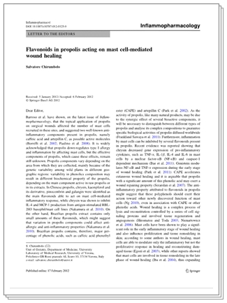
Flavonoids in propolis acting on mast cell-mediated wound healing
Inflammapharmacology, 2012 Feb 17
Salvatore Chirumbolo, University of Verona Italy 


Barroso et al. have shown, on the latest issue of Inflammopharmacology, that the topical application of propolis on surgical wounds affected the number of mast cells recruited in these sites, and suggested two well-known anti-inflammatory components present in propolis, namely caffeic acid and artepillin C, as possible active molecules (Borelli et al. 2002; Paulino et al. 2008). It is widely acknowledged that propolis down-regulates type I allergy and inflammation by affecting mast cells, but the effective components of propolis, which cause these effects, remain still unknown.

Propolis components vary depending on the area from which they are collected, mainly because of the genetic variability among wild plants in different geographic regions; variability in phenolics composition may result in different biochemical property of the propolis, depending on the main component active in raw propolis or in its extracts. In Chinese propolis, chrysin, kaempferol and its derivative, pinocembrin and galangin were identified as main flavonoids able to act on mast cell-mediated inflammatory response, while chrysin was shown to inhibit IL-4 and MCP-1 production from antigen-stimulated RBL-2H3 basophil/mast cell lines (Nakumura et al. 2010). On the other hand, Brazilian propolis extract contains only small amounts of these flavonoids, which might suggest that variation in propolis components could affect anti-allergic and anti-inflammatory properties (Nakumura et al. 2010). Brazilian propolis contains, therefore, major percentage of phenolic acids, such as caffeic acid phenethyl ester (CAPE) and artepillin C (Park et al. 2002). 

excerpt online at SpringerLink.com
As the activity of propolis, like many natural products, may be due to the synergic effect of several bioactive components, it will be necessary to distinguish between different types of propolis and analyse its complex compositions to guarantee specific biological activities of propolis diffused worldwide (Frankland Sawaya et al. 2011). Furthermore, inflammation by mast cells can be inhibited by several flavonoids present in propolis. Recent evidence was reported showing that chrysin decreased gene expression of pro-inflammatory cytokines, such as TNF-ά, IL-1β, IL-4 and IL-6 in mast cells by a nuclear factor-κB (NF- κ B) and caspase-1 dependent mechanism (Bae et al. 2011). Genistein modulates NF- κ  B and TNF-ά expression during the early stage of wound healing (Park et al. 2011). CAPE accelerates cutaneous wound healing and its is arguable that propolis with a significant amount of this phenolic acid may exert a wound repairing property (Serarslan et al. 2007). 

The anti-inflammatory property attributed to flavonoids in propolis might suggest that these polyphenols should exert their action toward other newly discovered function of mast cells (Ng 2010), even in association with CAPE or other phenolic acids. Wound healing is a complex process of lysis and reconstitution controlled by a series of cell signaling proteins and involved tissue regeneration and angiogenesis (Hiromatsu and Toda 2003; Nienartowicz et al. 2006). Mast cells have been shown to play a significant role in the early inflammatory stage of wound healing and also influence proliferation and tissue remodeling in skin…

Royal Jelly Protects Kidneys, Liver from Anti-Cancer Treatments

New research from Turkish researchers confirm the protective properties of Royal Jelly against damage caused by anti-cancer therapies. Could this be new ground towards complementary alternative therapies? 

Royal Jelly Modulates Oxidative Stress and Apoptosisin Liver and Kidneys of Rats Treated with Cisplatin

Abstract:
Cisplatin (CDDP) is one of the most active cytotoxicagents in the treatment of cancer and has adverse side effects such asnephrotoxicity and hepatotoxicity. The present study was designed to determinethe effects of royal jelly (RJ) against oxidative stress caused by CDDP injuryof the kidneys and liver, by measuring tissue biochemical and antioxidantparameters and investigating apoptosis immunohistochemically.

Twenty-four Sprague Dawley rats were divided into fourgroups, group C: control group received 0.9% saline; group CDDP: injected i.p.with cisplatin (CDDP, 7 mg kg(-1) body weight i.p., single dose); group RJ:treated for 15 consecutive days by gavage with RJ (300 mg/kg/day); group RJ +CDDP: treated by gavage with RJ 15 days following a single injection of CDDP.Malondialdehyde (MDA) and glutathione (GSH) levels, glutathione S-transferase(GST), glutathione peroxidase (GSH-Px), and superoxide dismutase (SOD)activities were determined in liver and kidney homogenates, and the liver andkidney were also histologically examined.

RJ elicited a significant protective effect towardsliver and kidney by decreasing the level of lipid peroxidation (MDA), elevatingthe level of GSH, and increasing the activities of GST, GSH-Px, and SOD. In theimmunohistochemical examinations were observed significantly enhanced apoptoticcell numbers and degenerative changes by cisplatin, but these histologicalchanges were lower in the liver and kidney tissues of RJ + CDDP group. Besides,treatment with RJ lead to an increase in antiapoptotic activity hepatocytes andtubular epithelium.

In conclusion, RJ may be used in combination withcisplatin in chemotherapy to improve cisplatin-induced oxidative stressparameters and apoptotic activity...

excerpt:
Recently, royal jelly (RJ) has received particularattention because of studies that have reported that it is a highly efficientantioxidant and has free radical scavenging capacity [4, 15]. Royal jelly is asecretion produced by the hypopharyngeal and mandibular glands of workerhoneybees (Apis mellifera). It contains many important compounds withbiological activity such as free amino acids, proteins, sugars, fatty acids,minerals, and vitamins [16]. So far, RJ has been demonstrated to possessseveral physiological activities in experimental animals, includingvasodilative and hypotensive activities [17], the induction of decrease inserum cholesterol levels [18], antimicrobial [19], antiallergic [20],anti-inflammatory [21], immunomodulatory [22, 23], and antioxidant properties[16]. In addition, Kanbur et al. [24] revealed the protective effect of RJagainst paracetamol-induced liver damage in mice.

Propolis Component Key in Treating Allergic Airway Disease


About this Journal

One of the many compounds of Propolis, CAPE has the super-hero force to aid the human condition...


Caffeic Acid Phenethyl Ester Suppresses Eotaxin Secretion and Nuclear p-STAT6 in Human Lung Fibroblast Cells
J Microbiol Immunol Infect, 2011 May 19

BACKGROUND: 

Caffeic acid phenethyl ester (CAPE), an active component of propolis, has been proven to have anti-inflammatory and antiallergic properties. We have investigated the activity of CAPE in regulating cytokine-induced eotaxin production and its related signal protein, signal transducer and activator of transcription 6 (STAT6), in human lung fibroblast.

METHODS: 

The CCD-11Lu human lung fibroblast cell line was used as an in vitro model. Cells were pretreated with CAPE followed by stimulation with interleukin-4 and tumor necrosis factor alpha. The levels of eotaxin in cultured supernatants were measured by enzyme-linked immunosorbent assay. The amounts of STAT6 and phosphorylated STAT6 in cellular nuclear protein extracts were determined by Western blot analysis. STAT6 DNA binding activities were detected by electrophoretic mobility shift assay.

RESULTS: 

Pretreated CCD-11Lu cells with noncytotoxic doses (0.1-10μM) of CAPE inhibited the production of eotaxin under stimulation of interleukin-4 (10ng/mL) and tumor necrosis factor alpha (10ng/mL). CAPE pretreatment also decreased the amount of phosphorylated STAT6 and the STAT6 DNA binding complexes in nuclear extracts.

CONCLUSION: 

CAPE inhibited the production of eotaxin protein in stimulated human lung fibroblast cells in a dose-dependent manner. This activity is, at least, through STAT6 inhibition. We suggest that CAPE is a promising agent in controlling eotaxin secretion and subsequent eosinophils influx and may therefore have a potential role to play in treating allergic airway disease
 
Support : Creating Website | SEO Template | Free Template
Copyright © 2011. Natural Medicine - All Rights Reserved
Proudly powered by Blogger