Showing posts with label pinocembrin. Show all posts

Propolis Flavonoids Accelerate Wound Healing

Propolis flavonoids are numerous but those of great interest are CAPE (caffeic acid phenethyl ester), chrysin, kaempferol, pinocembrin, galangin and artepillin C. These vary on the geographical source of propolis, due the wide variety of polyphenols harvested by the honeybees in the region. Nonetheless, the anti-inflammatory effects have great importance for many applications...

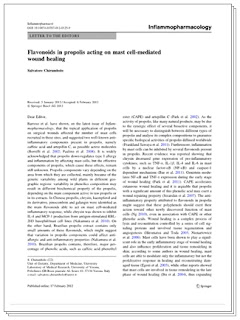
Flavonoids in propolis acting on mast cell-mediated wound healing
Inflammapharmacology, 2012 Feb 17
Salvatore Chirumbolo, University of Verona Italy 


Barroso et al. have shown, on the latest issue of Inflammopharmacology, that the topical application of propolis on surgical wounds affected the number of mast cells recruited in these sites, and suggested two well-known anti-inflammatory components present in propolis, namely caffeic acid and artepillin C, as possible active molecules (Borelli et al. 2002; Paulino et al. 2008). It is widely acknowledged that propolis down-regulates type I allergy and inflammation by affecting mast cells, but the effective components of propolis, which cause these effects, remain still unknown.

Propolis components vary depending on the area from which they are collected, mainly because of the genetic variability among wild plants in different geographic regions; variability in phenolics composition may result in different biochemical property of the propolis, depending on the main component active in raw propolis or in its extracts. In Chinese propolis, chrysin, kaempferol and its derivative, pinocembrin and galangin were identified as main flavonoids able to act on mast cell-mediated inflammatory response, while chrysin was shown to inhibit IL-4 and MCP-1 production from antigen-stimulated RBL-2H3 basophil/mast cell lines (Nakumura et al. 2010). On the other hand, Brazilian propolis extract contains only small amounts of these flavonoids, which might suggest that variation in propolis components could affect anti-allergic and anti-inflammatory properties (Nakumura et al. 2010). Brazilian propolis contains, therefore, major percentage of phenolic acids, such as caffeic acid phenethyl ester (CAPE) and artepillin C (Park et al. 2002). 

excerpt online at SpringerLink.com
As the activity of propolis, like many natural products, may be due to the synergic effect of several bioactive components, it will be necessary to distinguish between different types of propolis and analyse its complex compositions to guarantee specific biological activities of propolis diffused worldwide (Frankland Sawaya et al. 2011). Furthermore, inflammation by mast cells can be inhibited by several flavonoids present in propolis. Recent evidence was reported showing that chrysin decreased gene expression of pro-inflammatory cytokines, such as TNF-ά, IL-1β, IL-4 and IL-6 in mast cells by a nuclear factor-κB (NF- κ B) and caspase-1 dependent mechanism (Bae et al. 2011). Genistein modulates NF- κ  B and TNF-ά expression during the early stage of wound healing (Park et al. 2011). CAPE accelerates cutaneous wound healing and its is arguable that propolis with a significant amount of this phenolic acid may exert a wound repairing property (Serarslan et al. 2007). 

The anti-inflammatory property attributed to flavonoids in propolis might suggest that these polyphenols should exert their action toward other newly discovered function of mast cells (Ng 2010), even in association with CAPE or other phenolic acids. Wound healing is a complex process of lysis and reconstitution controlled by a series of cell signaling proteins and involved tissue regeneration and angiogenesis (Hiromatsu and Toda 2003; Nienartowicz et al. 2006). Mast cells have been shown to play a significant role in the early inflammatory stage of wound healing and also influence proliferation and tissue remodeling in skin…

Propolis Found to Inhibit Candida Albicans

The antifungal protection of Propolis is due to its rich, diverse flavonoid content and it's proven control of Candida Albicans has been validated in studies worldwide...

Structural and Genetic Alterations of Fungal Cells Caused by Mexican Propolis

In order to study the antifungal activity of propolis ethanol extracts (PEE) from Apis mellifera bees from Mexico we used optical microscopy and we were able to see that PEE induced inhibition of germ tube formation of C. albicans.


Ultrastructural findings were seen using transmission electron micrographs of ultrathin sections of Candida albicans revealed that these cells suffered vacuolization, increased formation of storage granules as well as alteration and disruption of the outer structures of yeast, with release of intracellular material. In order to describe the molecular mechanism of action, we evaluate gene expression...


We were able to identify some over- and under-expressed genes. ADH1 y PIK1 genes were under-expressed in a manner dependent on concentration and exposure time. On the other hand, our team has determined the chemical profile of several Mexican propolis, with interesting results as the diversity of content and different levels of antifungal and antibacterial activities


Although many studies have focused on demonstrating the antifungal activity of propolis and different extracts, few have shown their effects on the morphology and structure of fungi. We have found important cell damage caused by contact with extracts using bright field optical microscopy studies as well as the fluorescent dye White Calcofluor and transmission electron microscopy...


Based on the above results, it can be stated that propolis from the States of Guanajuato and Queretaro showed a higher number of compounds with longer retention time (higher polarity), which was corroborated by the MS study, in which it was found that these compounds were of the flavonoid-type, mainly pinocembrin, naringenin and chrysin, compounds characteristic of plant specimens visited by bees in that geographic locality...


In summary, we succeeded in establishing the chemical content of propolis, with a significant presence of flavonoid type compounds, a prerequisite for such high activity. Similarly, we must remember that the characteristic flora of each region determines the type of propolis bees collect in that locality, and this accounted for the differences in the antimicrobial activity which we found in this study.

Propolis Exhibits Cytotoxic Effect on Cervical Tumor Cells

Propolis always thrives in protecting the mucuous linings of the body from bacteria, viruses, fungi, inflammation and even tumor cell growth...

Chemical composition of the ethanolic propolis extracts and its effect on HeLa cells.


ETHNOPHARMACOLOGICAL RELEVANCE:
Propolis is a resinous hive product collected by honeybees from various plant sources. It is widely used in traditional medicine and is reported to have a broad spectrum of pharmacological effects (antibacterial, antihepatoxic, antioxidative, anti-inflammatory, etc.). Thus the aim of this study was to assess cytotoxic effect of various ethanol propolis extractions on the cervical tumor cell line (HeLa) and compare it with their phenolic acids and flavonoids composition.

MATERIALS AND METHODS:
Twenty samples of raw propolis were collected from 17 localities of Croatia (I-XVII), 2 of Bosnia and Hercegovina (XVIII, XIX) and 1 of Macedonia (XX). Reverse phase HPLC was used to determine the chemical composition of polyphenols. Biological experiments were carried out in vitro on cervix adenocarcinoma cell line (HeLa).

RESULTS:
Phenolic acids (ferulic acid, p-coumaric acid, caffeic acid) and flavonoids (tectochrysin, galangin, pinocembrin, pinocembrin-7-methylether, chrysin, apigenin, kaempferol, quercetin) have been determined using HPLC analysis in 20 ethanolic propolis extracts. All samples contain tectochrysin in ranges of 0.1988 mg/g (XVIII) to 1.2004 mg/g (III), while caffeic acid and quercetin have not been found. Flavonoid that is most abundant is galangin in ranges from 0.3706 mg/g (XVII) to 47.4879 mg/g (IX). The samples of propolis numbers I, VI and X applied in the investigated concentration range manifested significant reduction of cell growth. GI₅₀ value as indicator of cytotoxicity among propolis samples showed that propolis number VII is the most effective (GI₅₀=76 μg/ml) followed by propolis nos. XV, XVIII and I.

CONCLUSION:
Antiproliferative and cytotoxic effect of propolis on the HeLa cells is not correlating with the concentration of particular components but on establishing the possible synergistic antiproliferative activity of individual phenolic acid and flavonoids. Differences in the chemical composition lead to diversity in biological activity of propolis samples.
 
Support : Creating Website | SEO Template | Free Template
Copyright © 2011. Natural Medicine - All Rights Reserved
Proudly powered by Blogger